
Procedure map / Pain
Epiduroscopy
A thin endoscope passed through the catheter gives a direct view of the epidural space; the lesion is treated under vision.
Step by step
- 01
Access
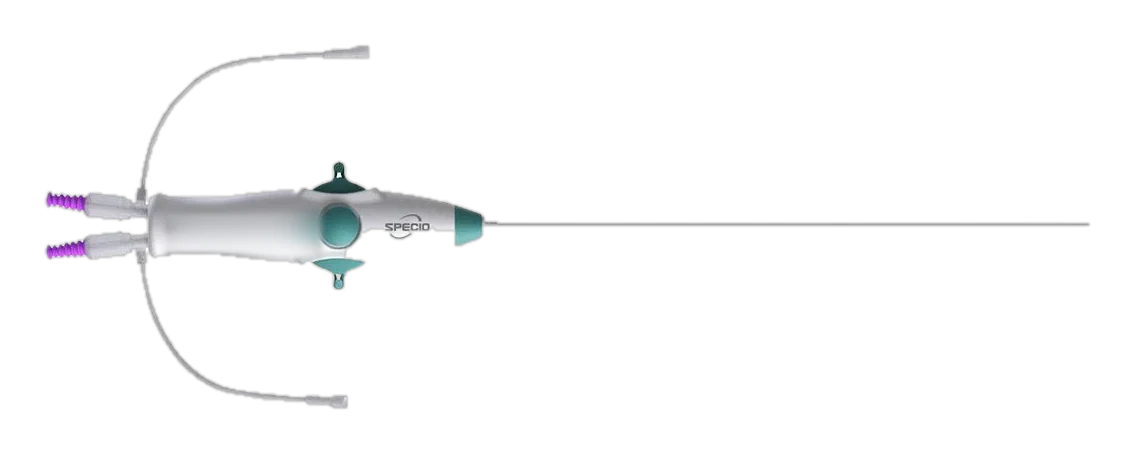
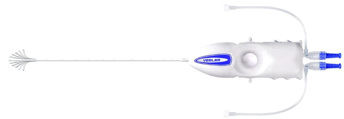
Under sedation the sacral hiatus is entered with a needle. The working path is opened with guide wire, dilator and sheath (Seldinger technique) or directly with a large-bore needle.
Two access kit options
- 02
Catheter and endoscope
A flexible endoscope is advanced through the catheter to the end of its hub and the catheter is filled with saline. The catheter is then advanced through the sheath or introducer to the target.
Outer diameter 3 mm, 2 working channels
- 03
Saline for visibility
Saline is given slowly through one channel to open the epidural space and clear the view. Volume and pressure are kept limited.
Dual infusion ports
- 04
Finding the lesion
The catheter tip is steered two ways through tight anatomy. Adhesions and inflamed nerve roots invisible on fluoroscopy are seen directly.
Two-way, locking tip
- 05
Treatment under vision
The adhesion is released with the catheter tip and medication is delivered to the target, all while watching the screen.
Medication is given through the injection port on the body
A general workflow is shown. Practice varies by physician and patient.